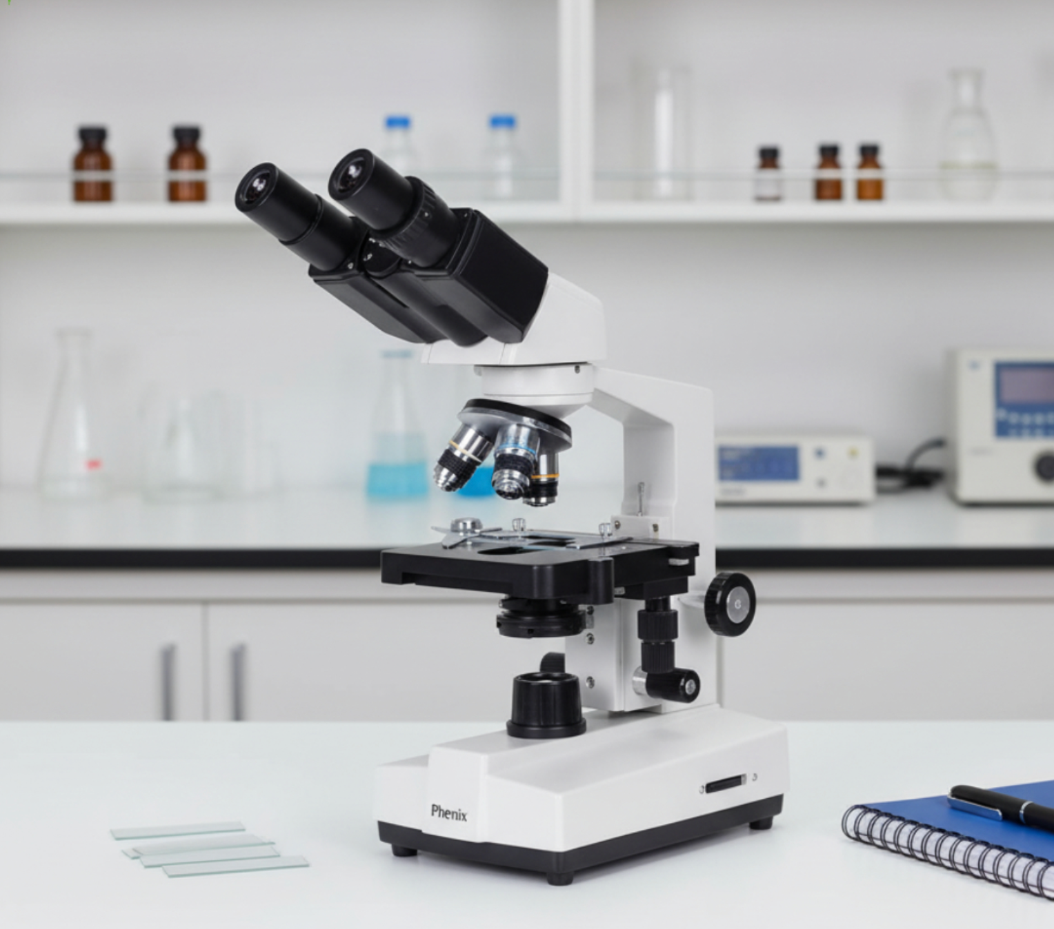
Description
Core Specifications & Optical Setup
-
Magnification Range: 40X to 1600X achieved via interchangeable eyepieces (typically WF10X and WF16X) paired with achromatic objectives (4X, 10X, 40X, and 100X oil immersion).
-
Illumination & Condenser: Built-in LED or halogen variable illumination system with an Abbe Condenser (N.A. 1.25) and iris diaphragm for optimal contrast tuning.
-
Focusing Mechanics: Coaxial or dual-layer coarse and fine focusing knobs for precise depth control at higher magnifications.
-
Stage: Double-layer mechanical stage with X-Y slide positioning for smooth specimen maneuvering.
Role in Medical Education
In medical schools, biology programs, and technical training labs, the XSP-36T serves as a primary teaching instrument due to its balance of simplicity and professional features:
-
Histology & Cytology Instruction: Perfect for teaching students how to identify tissue structures, cell membranes, epithelial layers, and organelle groupings.
-
Microbiology Fundamentals: With the 100X oil immersion objective (yielding 1000X–1600X total magnification), students learn slide preparation, Gram staining, and bacterial morphology identification (cocci, bacilli, spirilla).
-
Hematology Training: Enables clear visualization of blood smears, including red blood cell (RBC) counts, white blood cell (WBC) differential counting, and platelet observation.
Role in Research & Routine Laboratory Diagnostics
While high-end advanced research often utilizes infinity-corrected trinocular fluorescence or electron systems, the XSP-36T fills vital baseline functions in research and clinical settings:
-
Routine Sample Screening: Serves as a reliable workhorse for preliminary slide inspection before passing samples to specialized imaging units.
-
Parasitology & Urinalysis: Widely used in field clinics and entry-level research labs to detect intestinal parasites, ova, protozoa, sediment crystals, and fungal cultures.
-
Cell Culture Quality Control: Quick, routine checking of slide-mounted samples and tissue smears for contamination or general morphology checks.
Key Advantage: The XSP-36T provides true 1000X+ optical resolution required for viewing cellular and bacterial structures without the high acquisition or maintenance costs associated with complex digital research rigs.
Reviews
There are no reviews yet.